Hi Mariam - You have asked a good question!
Cells specialized to secrete protein hormones have a characteristic
appearance, reflecting the cellular machinery needed to synthesize and
secrete the protein hormones they produce. As an example, I'll include
an electron micrograph from a pituitary cell which secretes human
growth hormone. This picture was taken at *very* high magnifcation,
on the order of 10,000X! Rather than use light to 'see' the strctures
within the cell, electron microscopes (EM) use *electron beams* to visualize
micro-thin sections taken through cells. Each of the characteristic
organelles is labeled within the image.
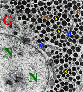 N -- Nucleus: The nucleus looks fairly normal
in this cell. Cells that produce lots of proteins require many ribosomes
to carry out the process of translation. Ribosomes are made both of
proteins, and ribosomal RNA - rRNA. rRNA is synthesized from the
corresponding rDNA in the nucleus, in a structured called the nucleolus.
Thus, secretory cells often have prominent nucleoli.
N -- Nucleus: The nucleus looks fairly normal
in this cell. Cells that produce lots of proteins require many ribosomes
to carry out the process of translation. Ribosomes are made both of
proteins, and ribosomal RNA - rRNA. rRNA is synthesized from the
corresponding rDNA in the nucleus, in a structured called the nucleolus.
Thus, secretory cells often have prominent nucleoli.
M -- Mitochondria: Mitochondira are the energy
factories of the cell. The regenerate spent ADP (adenosine di-phosphate)
into ATP (adenosine tri-phosphate), the energy currency of the cell.
Protein translation requires lots of energy, and thus lots of ATP, so
the cells needs to have lots of mitochondria to meet the demands.
ER -- (rough) Endoplasmic reticulum: Protein
translation takes place in the rough endoplasmic reticulum a
complex interconnecting maze of internal membranes supporting ribosomes
and the other cytoplasmic components needed to carry out protein translation.
It is called 'rough' as the presence of so many ribosomes gives it a
rough texture under EM. In contrast, smooth endoplasmic reticulum
has few to no ribosomes. Cells that secrete steroid hormones such
as testosterone, other androgens, and glucocorticoids have lots of
smooth ER. The enzymes responsible for synthesizing these non-protein
hormones do their thing in the smooth ER. You may find it interesting to
know that the structure of steroids closely resembles the structure of
cholesterol..
G -- Golgi: The Golgi body is the site of
protein glycosylation. Certain completed proteins are processed
through the Golgi so sugars/carbohydrates can be added on at specific
sites. This type of modification is called a post-translational
modification as the composition of the protein is altered
after translation has been completed. Enzymes called glycosyltransferases
(glyco = sugar; transferase = enzyme that 'transfers') add different
kinds of sugars. Sugars commonly added include mannose,
galactose, and fucose. They are added at specific sites
in the protein, always on residues of the amino acid asparagine.
The sugars often play important roles
in the functioning of the hormone. The sugars may make it more stable
when released into the blood, and they may also be necessary for it to
properly associate with the cell they intend to affect. This cell has a
well-developed Golgi apparatus.
S -- secretory granule Lastly, hormones are
packaged into secretory granules, probably the most prominent feature
in the image. These small vesicles store the hormone until it is ready to
be released, at Which point the vesicles fuse with the plasma membrane of
the cell to release their contents.
Hope this helps..
-NB
Current Queue |
Current Queue for Cell Biology |
Cell Biology archives
Return to the MadSci Network
MadSci Home | Information |
Search |
Random Knowledge Generator |
MadSci Archives |
Mad Library | MAD Labs |
MAD FAQs |
Ask a ? |
Join Us! |
Help Support MadSci
MadSci Network
© 1997, Washington University Medical School
webadmin@www.madsci.org