Re: Why does the right side of the heart have a TRICUSPID valve?
Area: Anatomy
Posted By: Lynn Bry, MD/PhD Student, Washington University Medical School
Date: Mon Feb 17 22:43:47 1997
Message ID: 855255174.An
> Why does the right side of the heart have a TRICUSPID valve?
As far as I have been able to find, there is (currently) no 'definitive'
answer as to why a three-leafed tricuspid valve separates the right
atrium from the left, and a two-leafed bicuspid valve (the mitral valve)
separates the left atrium from the left ventricle. However, I can say
two things with certainty:
- The different valves arise over the course of fetal development.
- Spatial or 'topological' arrangements of developing
heart tissue appear to influence how the valves form, and whether
two or three leaflets will develop on a particular side of the heart.
If you're impatient, you can look at the pictures then
jump to the conclusion.
 SOME BACKGROUND
The embryology of the heart is incredibly complicated. However, I think
it would suffice to say that we don't start out with a 4 chambered heart.
Within the first 3-4 weeks of human development a single large vessel
undergoes a series of rotations to create the primitive atria which
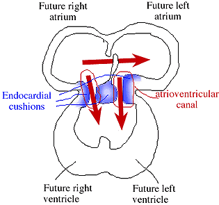
connect to a single ventricle. The first figure shows a cross-section
through the heart at this stage. Note that a 'flap valve' separates the
two atria (the foramen ovale, or oval opening). While in the womb,
blood is oxygenated within the placenta - a rich nexus of
blood vessels where the fetal circulation exchanges oxygen and receives
nutrients from the maternal circulation. Exchange occurs in the placenta as
the lungs do not function until birth. Thus the heart must shunt
blood away from the lungs. It accomplishes this in a variety of ways.
- Blood entering the right atrium is shunted to the left atrium
across the foramen ovale. In utero, pressures in the right side of the heart
are greater than in the left side. Blood readily flows from right ->
left. Once the lungs fill with air, however, the situation reverses.
Pressures on the left side are then greater than on the right. By the time of
birth the connections between the right and left side have closed (or
so one hopes..), so there is no flow of blood from left to right.
- Blood making it into the right ventricle can readily cross into the
left ventricle as the interventricular septum has not yet formed to
separate the right ventricle from the left ventricle.
- (Not shown). Blood entering the Pulmonary arteries from
the right ventricle can cross directly into the aorta via a
connection called the ductus arteriosis. This connection normally
closes a few days after birth.
Aside from having a single ventricle, you can see that nothing separates the
atria from the ventricles. They form a continuous canal - the
atrioventricular canal. Within this area lie the endocardial
cushions (shown in blue), areas of developing tissue that form part of
the ventricular walls. Most people believe they contribute little to
no tissue to the actual valve leaflets. However, their position
within the atrioventricular canal is of importance. They also fuse during the
4th week of development to form a ring of tissue from which the valves will
develop.
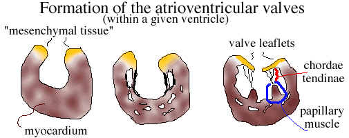 The second figure shows how the atrioventricular valves develop. The figure
cuts through an 'ideal ventricle.' 'Mesenchymal tissue' (yellow) near
the area of the AV canal will form the valve leaflets. Over time the
vetricular tissue is 'hollowed out' so the mesenchymal tissue overhangs
the receding myocardium. Degenerating ventricular muscle forms the
chordae tendineae - thick stringy attachments which connect the
valve leaflets to the papillary muscles of the ventricular wall.
These muscles contract with the rest of the ventricle when
blood is being moved into either the aorta or the pulmonary vessels.
Without their contraction, the pressure of the blood could force the tricuspid and
mitral valve leaflets back into the atrial chamber (a bad thing..)
The second figure shows how the atrioventricular valves develop. The figure
cuts through an 'ideal ventricle.' 'Mesenchymal tissue' (yellow) near
the area of the AV canal will form the valve leaflets. Over time the
vetricular tissue is 'hollowed out' so the mesenchymal tissue overhangs
the receding myocardium. Degenerating ventricular muscle forms the
chordae tendineae - thick stringy attachments which connect the
valve leaflets to the papillary muscles of the ventricular wall.
These muscles contract with the rest of the ventricle when
blood is being moved into either the aorta or the pulmonary vessels.
Without their contraction, the pressure of the blood could force the tricuspid and
mitral valve leaflets back into the atrial chamber (a bad thing..)
THE ANSWER
As I said, eariler, the position of the endocardial cushions
was of importance. It is believed the cushion tissue shown in the middle
of Figure 1 bulges more on the left side of the heart than on the right
side. Some studies suggest this bulge becomes incorporated into the mitral
valve by fusing two areas of the valvular ring of tissue
into a single leaflet. The remaining tissue develops into the second
valve leaflet (a bicuspid valve). On the right side of the heart, there is
no similar bulge, so the developing valvular tissue splits into three
leaflets -> the tricuspid valve.
REFERENCES:
Wenink & Gittenberger-de Groot: Embryology of the Mitral Valve
Int. J. of Cardiology, 11: 75-84, 1986.
Langman's Medical Embryology, 6th ed. 1990
ISBN - 0-683-07493-8
Current Queue |
Current Queue for Anatomy |
Anatomy archives
Return to the MadSci Network
MadSci Home | Information |
Search |
Random Knowledge Generator |
MadSci Archives |
Mad Library | MAD Labs |
MAD FAQs |
Ask a ? |
Join Us! |
Help Support MadSci
MadSci Network
© 1997, Washington University Medical School
webadmin@www.madsci.org