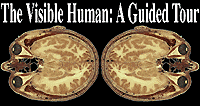

This site illustrates two low-tech, simple ways to create annotated images (images associating textual information with graphic elements) by extrapolating upon common imagemaps, or by using javascripts. Such images have a particular educational appeal as they let the user focus on specific information within an image. They also provide a handy means to allow pathologists to include text-based comments with digitized pathology images.
The "Visible Human" site presents anatomical information to a lay audience, while the "Flow Cytometry Archive" illustrates a more sophisticated interface used by the pathology residents and staff at Brigham & Women's hospital. Click on the images below to sample these sites.

|

|
Return to the Visible Human, A Guided Tour | The MAD Scientist Network